Gamma Knife Surgery: What It Is, How It Works, and Who Needs It

None of that is accurate. Gamma Knife surgery involves no knife whatsoever. No incision. No opening of the skull. No general anesthesia in most cases. No hospital stay overnight.
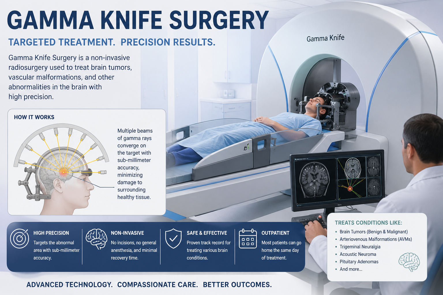
What it does involve is 192 precisely focused beams of radiation converging simultaneously on a single point inside the brain with extraordinary accuracy — and that convergence destroys tumor tissue without a surgeon’s hand ever entering the skull.
When my uncle was told he needed Gamma Knife treatment for a small meningioma, he spent a week assuming he was facing brain surgery. Understanding what Gamma Knife actually involves changed everything about how he approached it.
What Gamma Knife Actually Is
Gamma Knife is a form of stereotactic radiosurgery — a technique that uses multiple radiation beams focused with extreme precision on a specific target in the brain.
The Gamma Knife system uses 192 individual cobalt-60 radiation sources arranged in a helmet-shaped device. Each source emits a single focused beam of gamma radiation. On its own, each beam carries too little energy to damage tissue it passes through. All 192 beams converge precisely on a single predetermined point — the tumor target. At that convergence point, the combined radiation dose is high enough to destroy tumor cells.
The result is tumor tissue damage contained almost entirely within the target volume, with rapid dose falloff in surrounding normal brain. Structures just millimeters away receive a tiny fraction of the tumor dose.
Swedish neurosurgeon Lars Leksell invented the Gamma Knife in 1967. The technology has evolved continuously since then, with the current Icon system adding real-time imaging guidance throughout treatment for even greater precision.
What Tumors Gamma Knife Treats
Gamma Knife works best for small, well-defined targets — generally tumors up to approximately 3 to 4 centimeters in diameter. Larger tumors or tumors with irregular borders are less suited to this approach.
Brain metastases represent the most common use of Gamma Knife worldwide. Cancer that has spread to the brain from lung, breast, kidney, colon, or skin can be treated with Gamma Knife — often multiple lesions in a single session. It provides excellent local control with minimal disruption to surrounding brain.
Meningiomas respond well to Gamma Knife, particularly smaller tumors and those in surgically challenging locations — near the cavernous sinus, optic nerve, or brainstem. Control rates for Grade 1 meningiomas exceed 90% over five years.
Acoustic neuromas — benign tumors on the hearing nerve — are a classic Gamma Knife target. The treatment controls tumor growth in over 90% of cases while preserving hearing function in many patients.
Pituitary adenomas that have regrown after surgery, or functioning tumors producing excess hormones, respond to Gamma Knife with good control rates.
Arteriovenous malformations (AVMs) — abnormal tangles of blood vessels — obliterate slowly over two to three years after Gamma Knife treatment, eliminating bleeding risk.
Recurrent gliomas in selected patients can receive Gamma Knife as part of retreatment approaches, though results in infiltrating gliomas are less predictable than in well-defined targets.
Trigeminal neuralgia — severe facial pain caused by nerve compression — responds to Gamma Knife targeting of the trigeminal nerve root, providing pain relief in most patients.
How Gamma Knife Differs From Conventional Surgery
Conventional surgery physically removes tumor tissue. A neurosurgeon opens the skull, enters the brain, and excises tumor cells directly. This approach works best for large tumors causing symptoms from mass effect and for tumors in accessible locations where complete removal is achievable.
Gamma Knife doesn’t remove anything. It damages tumor cells so severely that they can no longer reproduce. The treated cells die over days to months. The dead tissue gradually shrinks on MRI scans over months to years after treatment. The tumor doesn’t disappear immediately — it dies slowly.
Neither approach is universally superior. Large tumors causing significant brain compression need physical removal. Small, deep tumors in surgically risky locations — where a craniotomy would cause significant damage — are often better suited to Gamma Knife. Some tumors benefit from both: surgery to remove the bulk, then Gamma Knife to address residual tumor.
The Gamma Knife Treatment Day — Step by Step
Arrival and Frame Placement
Patients arrive in the morning on treatment day. The procedure begins with frame placement — a stereotactic head frame attaches to the skull using four small pins inserted under local anesthesia. This frame provides the coordinate system that guides radiation delivery with sub-millimeter accuracy.
Frame placement is the part patients most commonly describe as uncomfortable. The pins create brief stinging during local anesthetic injection. Once numb, most patients feel pressure rather than pain during actual pin placement. The frame remains in place throughout the entire treatment day.
Frameless systems — using a custom mask rather than a pin-based frame — are now available at some centers and eliminate the frame-placement step entirely.
Imaging
After frame placement, high-resolution MRI with contrast provides the detailed brain images used for treatment planning. In some cases, CT or angiography adds additional information.
Treatment Planning
A team of neurosurgeons, radiation oncologists, and medical physicists reviews the images together. Using specialized software, they define the target volume precisely and design the radiation plan — selecting shot sizes, positions, and timing to cover the target while minimizing dose to surrounding structures.
Planning takes one to several hours depending on target complexity.
Treatment
The patient lies on the treatment table. The head frame connects to the Gamma Knife helmet. The table slides into the machine, positioning the target exactly at the focal point of the 192 beams.
Treatment itself is completely painless. Patients lie still. The machine makes quiet sounds. Some patients sleep. Others listen to music. The team monitors via camera throughout and can stop treatment instantly if needed.
Treatment duration ranges from 15 minutes to several hours depending on target size, shape, and prescribed dose. Multiple targets can be treated sequentially in the same session.
Discharge
After treatment, the frame removes — again with local anesthesia at the pin sites. Patients rest briefly. Most go home the same day. The small pin sites heal within days.
What to Expect After Gamma Knife
Immediate Aftermath
Fatigue on treatment day is common. Most patients rest at home that evening. Many return to work within one to two days. Driving is typically prohibited for 24 hours after the procedure.
Pin site discomfort resolves within days. Mild headaches occur in some patients and respond to standard pain relievers.
In the Weeks and Months After
Gamma Knife results aren’t immediate. The treated tumor doesn’t disappear from the next MRI scan. Instead, it may show various changes over the following months — swelling initially, then gradual shrinkage over one to three years as dead cells clear.
Follow-up MRI scans — typically at three, six, and twelve months after treatment, then annually — track the tumor’s response. Radiologists and oncologists interpret these changes in context. A tumor that appears to grow slightly in the first few months may be showing radiation swelling rather than true growth.
Distinguishing treatment effect from true tumor growth can require specialized MRI sequences, PET scanning, or clinical observation over time. This uncertainty during the monitoring period is one of the genuinely difficult aspects of Gamma Knife follow-up.
Effectiveness
Local tumor control rates for appropriately selected targets are excellent. Brain metastases show local control rates of 80% to 95% at one year. Meningiomas and acoustic neuromas show control rates exceeding 90% at five years. These are among the best outcomes in brain tumor treatment.
Side Effects and Risks
Gamma Knife carries a significantly lower complication rate than open craniotomy. However, radiation effects are real and can cause problems.
Radiation swelling — edema in and around the treated area — develops in some patients weeks to months after treatment. It can worsen neurological symptoms temporarily. Corticosteroids manage significant swelling effectively. Most swelling resolves with time.
Radiation necrosis — breakdown of brain tissue that received high radiation doses — occurs in a small percentage of patients, typically months to years after treatment. It can mimic tumor recurrence on MRI and occasionally causes significant neurological symptoms requiring treatment.
Hearing effects — relevant for acoustic neuroma treatment. Gamma Knife preserves serviceable hearing in approximately 50% to 70% of patients. This rate improves with lower doses but must be balanced against tumor control rates.
Hormonal effects from pituitary treatment can take months to years to appear and require long-term monitoring.
Is Gamma Knife Right for You?
Several factors determine whether Gamma Knife is appropriate for a specific patient and tumor. Tumor size matters — most centers limit Gamma Knife to targets under 3 to 4 centimeters. Location matters — targets near critical structures require careful planning but are often excellent Gamma Knife candidates precisely because surgery there carries high risk. Number of lesions matters for brain metastases — radiosurgery is generally preferred for patients with four or fewer metastases.
Overall health, prior radiation history, and the tumor’s molecular characteristics all influence decisions. Multidisciplinary tumor board review considers all these factors when recommending between surgery, Gamma Knife, conventional radiation, and combinations.
Questions to Ask About Gamma Knife
- Is my tumor’s size and location suitable for Gamma Knife?
- Will I need a head frame, or is frameless treatment available here?
- How many treatment sessions will I need?
- How will we distinguish radiation swelling from tumor recurrence on follow-up MRI?
- What is the expected control rate for my specific tumor type?
- Are there risks specific to my tumor’s location I should understand?
- Would surgery followed by Gamma Knife be better than Gamma Knife alone?
A Final Word
My uncle’s Gamma Knife treatment took about four hours total — frame placement, imaging, planning, and treatment combined. He went home that afternoon. He was back to his normal routine within two days.
His meningioma has remained stable through three years of follow-up MRI scans. He checks in with his radiation oncologist annually. He barely thinks about the tumor between appointments.
That outcome — stable tumor, normal life, minimal disruption — is exactly what appropriately selected Gamma Knife candidates achieve regularly. Understanding what the treatment actually involves replaced weeks of unnecessary fear with appropriate preparation.
The name is misleading. The reality is far more manageable than it sounds.
For more information about Gamma Knife and stereotactic radiosurgery, visit the
Mayo Clinic Gamma Knife Radiosurgery page or the
National Brain Tumor Society Radiation Therapy resource.
Disclaimer: This article serves educational and informational purposes only. It does not constitute medical advice. Please consult a qualified neurosurgeon or radiation oncologist for guidance specific to any individual diagnosis.