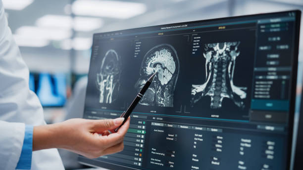
What Is a Brain MRI and When Should You Get One?
I’m not claustrophobic. But I understood in that moment why some people are genuinely distressed by the experience — especially when they’re already anxious about what the scan might show.
That experience made me want to write the guide I wish I’d had beforehand. Not just what an MRI is technically, but what it actually feels like, why doctors order it, what happens with contrast, what it can and can’t show, and most importantly — when you should be pushing to get one rather than waiting.
What Is a Brain MRI?
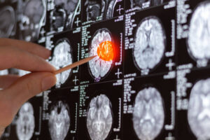
image of human brain. Medical treatmant concept. Diagnosis of the disease humans head. Close up.
MRI stands for Magnetic Resonance Imaging. Despite the intimidating name, the core concept is actually straightforward once you strip away the jargon.
Your body contains an enormous number of hydrogen atoms — particularly in water and fat. An MRI machine uses a powerful magnetic field combined with radio waves to temporarily align those hydrogen atoms and then measure how they respond when the magnetic field is briefly disrupted. Different types of tissue respond differently, and a computer converts those differences into extraordinarily detailed images.
The result is a three-dimensional picture of the brain’s soft tissue in extraordinary detail — far more detail than X-rays or CT scans can provide for this type of tissue. An MRI can show the brain’s structure in multiple planes: horizontal slices from top to bottom, vertical slices from front to back, and side-to-side slices from left to right, all from a single scan session.
Crucially, MRI uses no ionizing radiation. Unlike X-rays and CT scans, which pass radiation through the body to create images, MRI achieves its results entirely through magnetic fields and radio waves. This is one of the reasons it’s the preferred imaging tool for the brain, and why it can be used more freely in younger patients or in people who need repeated imaging over time.
Brain MRI vs CT Scan — What’s the Difference?

This is probably the most common question people ask when they’re first told they need brain imaging, so it’s worth addressing directly.
A CT scan — Computed Tomography — uses X-rays taken rapidly from multiple angles and processed by a computer to create cross-sectional images. It’s fast, usually taking only a few minutes, and it’s widely available even in smaller hospitals and emergency departments.
A brain MRI typically takes 30 to 60 minutes, requires a larger and more specialized machine, and is not available in every facility. But in terms of detail for soft tissue — which is what brain tissue is — MRI is significantly superior. It can detect smaller lesions, distinguish between different types of tissue more accurately, and visualize areas of the brain that are difficult to see clearly on CT due to bone interference.
In practice, the two tests are often used in sequence. A CT scan is ordered first in emergency situations because speed matters — it’s fast and can quickly rule out major bleeding, large tumors, or significant swelling. Once the immediate picture is established, an MRI follows to get the detailed assessment that CT alone can’t provide.
If a doctor suspects a brain tumor specifically, MRI is the test they actually want. CT is often a practical first step, not the final answer.
Brain MRI With Contrast vs Without Contrast
When a brain tumor or other serious pathology is suspected, the MRI is almost always ordered “with contrast” — meaning with a contrast agent injected into the bloodstream before or during the scan.
The contrast agent most commonly used is called gadolinium. It’s a metallic element that accumulates in areas where the blood-brain barrier has been disrupted or where blood flow is abnormally high — both of which are characteristic of many tumors and inflammatory conditions.
On the resulting images, areas where gadolinium has accumulated appear significantly brighter than surrounding tissue. This “enhancement” helps radiologists identify lesions that might be less visible on a non-contrast scan, understand their borders more clearly, and get a better sense of their likely nature.
For most people, gadolinium contrast is well-tolerated. Some people notice a brief sensation of warmth or a metallic taste in the mouth when it’s injected, both of which pass quickly. Allergic reactions are uncommon but not impossible, which is why you’ll be asked about prior reactions to contrast agents before the injection.
People with significantly reduced kidney function may not be able to receive gadolinium safely, since the kidneys are responsible for clearing it from the body. If kidney function is a concern, your doctor will check relevant blood tests before ordering contrast MRI.
What Actually Happens During a Brain MRI
Understanding what the experience involves makes it significantly less intimidating. Here’s exactly what to expect.
Before the Scan
You’ll be asked to remove all metal objects — jewelry, hair clips, piercings, watches, and anything else containing metal. You’ll also be asked about any metal implants in your body, including surgical plates or screws, cochlear implants, certain heart valves, and any embedded metal fragments. The MRI’s powerful magnetic field can interact dangerously with certain metal implants, so this screening is taken seriously.
Most pacemakers are now MRI-conditional rather than absolutely contraindicated, but this needs to be verified with the specific device’s documentation before proceeding.
You’ll change into a hospital gown and be given a locker for your belongings. If you’re having contrast MRI, an IV line will be placed in your arm — usually a simple process that takes a minute or two.
During the Scan
You’ll lie on a narrow table that slides into the center of the MRI machine. For a brain MRI, your head is positioned inside a frame called a head coil, which looks a bit like a cage surrounding your face and head. This coil is what receives the signals from your brain tissue — it doesn’t move or compress, it simply sits around your head.
Earplugs or headphones are provided because the machine is genuinely loud. The banging, knocking, and buzzing sounds are produced by the rapid switching of the magnetic gradients inside the machine — this is normal and expected, not a sign that anything is wrong.
The most important requirement is staying completely still. Even small movements can blur the images, potentially requiring sequences to be repeated and extending the total scan time. If you’re genuinely anxious about your ability to stay still, tell the technologist beforehand — they can position you more comfortably and some facilities can offer mild sedation if needed.
Throughout the scan, you’ll be able to hear the technologist through an intercom, and you’ll have a call button to press if you need to stop for any reason.
If You’re Claustrophobic
This is a real and valid concern, and it’s worth raising before your appointment rather than discovering it’s a problem when you’re already on the table.
Standard MRI machines have a bore — the central tunnel — that is typically about 60 to 70 centimeters wide. For some people, the combination of the narrow space and the noise creates genuine distress.
Options include asking your doctor about anti-anxiety medication taken beforehand, requesting a “wide-bore” MRI machine which has a larger opening, or in some cases using an “open MRI” which has a different configuration without an enclosed tunnel. Open MRI machines produce somewhat lower-quality images but are a reasonable alternative for people with significant claustrophobia.
After the Scan
Once the scan is complete, the table slides out, the IV line is removed if contrast was used, and you’re free to go. There’s no recovery time needed, no activity restrictions, and no side effects from the magnetic field itself.
The images go to a radiologist — a doctor who specializes in interpreting medical imaging — who analyzes them and produces a written report. Depending on the urgency of your situation and how the facility operates, results may be available to your referring doctor within hours or may take a few days.
What Can a Brain MRI Detect?
Brain MRI is an extraordinarily versatile diagnostic tool. Beyond brain tumors, it can detect and evaluate a wide range of conditions.
Brain tumors — both primary tumors that originate in the brain and metastatic tumors that have spread from cancer elsewhere in the body.
Stroke — particularly diffusion-weighted MRI sequences are highly sensitive for detecting acute stroke, sometimes within minutes of onset.
Multiple sclerosis — the characteristic lesions of MS appear clearly on MRI, and tracking them over time helps monitor disease progression.
Brain aneurysms — abnormal bulging in blood vessel walls that carries a risk of rupture.
Bleeding in or around the brain — including subdural hematomas, epidural hematomas, and intracerebral hemorrhage.
Infections — brain abscesses, encephalitis, and meningitis can produce characteristic findings on MRI.
Hydrocephalus — abnormal buildup of cerebrospinal fluid causing enlarged ventricles in the brain.
Dementia evaluation — MRI can show patterns of brain atrophy consistent with Alzheimer’s disease and other dementias, helping with diagnosis and monitoring.
Epilepsy investigation — certain structural abnormalities that cause seizures can be identified on dedicated epilepsy-protocol MRI sequences.
Pituitary gland abnormalities — including pituitary tumors that may be affecting hormone production.
When Should You Ask for a Brain MRI?
This is the question most people really want answered, and it deserves a direct response rather than vague hedging.
You should talk to a doctor about whether a brain MRI is appropriate if you are experiencing any of the following situations.
Headaches That Are New and Different
If you’ve developed a headache pattern that is new for you — not your typical tension headache, but something that feels different in quality or location — particularly if those headaches are progressively worsening over weeks or months, that warrants evaluation. The specific patterns most concerning are headaches that are worst in the morning, headaches that wake you from sleep, or headaches that worsen with bending forward, coughing, or straining.
A First Seizure
Any first-time seizure in an adult with no prior epilepsy diagnosis requires brain imaging. Full stop. Whether it was a dramatic generalized convulsion or a brief, subtle focal episode, the cause needs to be identified.
Unexplained Vision Changes
If your optometrist has examined your eyes and cannot explain your vision symptoms, or if your symptoms don’t improve with corrected lenses, a neurological cause including a brain issue should be investigated.
Sudden or Progressive Neurological Symptoms
Sudden weakness or numbness on one side of the body. Sudden severe headache unlike any you’ve had before. Sudden loss of speech or difficulty understanding language. Any of these warrants emergency evaluation immediately, not a scheduled appointment.
Progressive symptoms that develop over weeks or months — gradually worsening balance, steadily increasing cognitive difficulties, slowly progressive weakness — are less immediately emergency-level but still require investigation without significant delay.
Personality or Cognitive Changes
If someone close to you has undergone noticeable personality changes, significant memory decline, or behavioral changes that don’t have an obvious psychological or social explanation, neurological evaluation including brain imaging may be appropriate.
Monitoring a Known Condition
If you’ve already been diagnosed with a brain tumor, MS, a prior stroke, or another condition that requires follow-up, your neurologist or oncologist will have you on a scheduled MRI monitoring program. These follow-up scans are just as important as the initial diagnostic one.
Family History of Brain Aneurysm
People with a close family member who has had a brain aneurysm — particularly if multiple family members are affected — may be candidates for screening MRI angiography to look for aneurysms before they cause problems. This is a conversation worth having with your doctor.
What a Brain MRI Cannot Do
It’s worth being honest about the limitations, because MRI is sometimes spoken about as if it shows everything with perfect certainty.
MRI cannot always distinguish with certainty between different tumor types — tissue biopsy is still required for definitive diagnosis in most cases. It cannot reliably detect very small lesions below a certain size. Imaging artifacts — distortions created by movement, metal implants, or other factors — can occasionally make interpretation more difficult. And MRI shows structure, not always function — how the brain is working is a different question from what the brain looks like.
A normal brain MRI is genuinely reassuring, but it doesn’t absolutely guarantee that nothing is wrong. If symptoms persist after a normal MRI, continued follow-up with a neurologist remains appropriate.
A Final Word
Going back to that first MRI experience — the noise, the stillness, the strange intimacy of having a machine create an image of the inside of your skull — what I remember most is how the radiographer spoke to me beforehand.
She explained exactly what the sounds would be, approximately how long each sequence would take, and what would happen with the contrast. It took her maybe four minutes to explain. Those four minutes made the difference between an anxious thirty-five minutes and a manageable thirty-five minutes.
Information does that. It doesn’t change what the scan shows. But it changes how you experience the waiting, the process, and whatever comes next.
If you’re heading toward a brain MRI — whether because of something a doctor found or because you’re pushing to get one yourself — you now know more than I did going in. And that’s worth something.
For more information, visit the
Mayo Clinic Brain Tumor Guide or the
American Cancer Society.
Disclaimer: This article is written for educational and informational purposes only and does not constitute medical advice. If you are experiencing neurological symptoms, please consult a qualified healthcare professional promptly. MRI protocols and availability vary between medical facilities and countries.