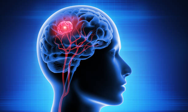
Brain Tumor Surgery: A Complete Guide for Patients and Families
Those questions kept her awake. And in my experience, they keep most families awake the night before brain surgery.
This guide answers every one of them. Not to replace the conversation with your surgical team — but to make that conversation more productive, and to give patients and families a real understanding of the process before, during, and after the operation.
Why Surgery Is Usually the First Step
Brain tumor surgery serves multiple purposes simultaneously. It removes tumor tissue — reducing pressure on the brain and improving symptoms immediately. It provides tissue for pathology and molecular testing, which determines the exact tumor type, grade, and genetic characteristics. That information shapes every treatment decision that follows surgery.
Surgery also reduces the total tumor burden — the amount of tumor remaining in the brain. Radiation and chemotherapy work better when they have less tumor to control. Even partial removal of a large, aggressive tumor significantly improves the effectiveness of subsequent treatment.
Not every brain tumor requires immediate surgery. Small, slow-growing, asymptomatic tumors sometimes call for active surveillance instead. But when surgery is recommended, understanding what it involves makes the entire experience less frightening and the recovery more manageable.
Types of Brain Tumor Surgery
Craniotomy — The Most Common Approach
A craniotomy is the standard surgical procedure for accessing and removing a brain tumor. The word means exactly what it sounds like: opening the cranium.
The neurosurgeon removes a section of the skull bone — called a bone flap — to access the brain beneath. After completing the tumor removal, the surgeon replaces and secures the bone flap precisely where it came from. Titanium plates and screws hold it in position while it heals back into place over the following weeks.
The size and location of the bone flap depends entirely on where the tumor sits. A tumor near the surface requires a smaller opening than one buried deeper in the brain. A tumor near the back of the head requires a differently positioned craniotomy than one near the front.
Awake Craniotomy
Awake craniotomy sounds alarming to most people hearing about it for the first time. The reality is more manageable than it sounds.
Surgeons use this technique when a tumor sits near areas of the brain controlling speech, language, or movement. Removing tumor tissue in these areas risks damaging functions the patient cannot afford to lose. Operating while the patient is conscious — and responsive — allows real-time monitoring of exactly which tissue performs which function.
During an awake craniotomy, the patient receives sedation and local anesthesia throughout the procedure. The brain itself has no pain receptors, so patients feel no pain from the surgical work on brain tissue. At specific moments during surgery, the anesthesiologist reduces sedation enough for the patient to respond to questions, follow simple commands, or read words aloud.
As the surgeon approaches tissue near critical functional areas, they use gentle electrical stimulation to temporarily disrupt function in small areas. If stimulating a specific spot causes the patient to stop speaking or lose movement in a limb, the surgeon knows that spot controls speech or movement and must be preserved. Tissue that can be removed safely shows no functional response to stimulation.
Patients who have undergone awake craniotomy frequently describe the experience as far less distressing than they anticipated. Careful preparation, good communication with the surgical team, and the genuine commitment to preserving function make the procedure achievable for the vast majority of appropriate candidates.
Stereotactic Needle Biopsy
When a tumor cannot be safely resected — due to its location deep in the brain, near critical structures, or in multiple locations — a stereotactic needle biopsy provides tissue for diagnosis without attempting removal.
The surgeon drills a small hole in the skull and inserts a thin needle guided by three-dimensional coordinates mapped from the pre-operative MRI. The needle collects a small sample of tumor tissue. The hole requires no plate or major repair — it heals naturally.
Most patients stay in hospital for one to two days after a stereotactic biopsy and recover quickly. The tissue obtained goes to pathology for the same detailed analysis performed on tissue from full resection.
Endoscopic Surgery
Endoscopic brain surgery uses a thin tube — called an endoscope — inserted through a small opening in the skull or through a natural body passage. A tiny camera at the tip transmits images to a monitor, guiding the surgeon’s instruments.
Surgeons use endoscopic approaches primarily for tumors in specific locations: pituitary tumors accessed through the nasal cavity and sinus passages, cysts within the brain’s fluid-filled ventricles, and certain other tumors in locations where a traditional craniotomy would cause more damage than the endoscopic approach.
Endoscopic surgery typically produces faster recovery and less disruption to surrounding structures than open craniotomy, when tumor location makes it appropriate.
Laser Interstitial Thermal Therapy (LITT)
LITT is a minimally invasive technique gaining increasing use for brain tumors in locations too deep or risky for standard surgery. The surgeon drills a small hole in the skull and inserts a thin laser probe directly into the tumor under MRI guidance. The laser heats and destroys tumor tissue from within, while real-time MRI monitoring allows the surgeon to watch the heat spreading through the tumor and stop precisely when the target area is treated.
Patients recover faster from LITT than from open craniotomy. Hospital stays of one to two days are typical. The approach works particularly well for small tumors, radiation necrosis, and tumors in deep brain regions where open surgery carries high risk.
Before Surgery: Preparation
Pre-Operative Testing
The surgical team orders several tests before the operation. Blood tests establish baseline values and check clotting function — abnormal clotting increases surgical bleeding risk. An electrocardiogram checks heart function. Chest X-ray evaluates lung health. Additional brain imaging — often a specialized pre-operative MRI — maps the tumor’s precise relationship to critical brain structures, blood vessels, and functional areas.
Functional Mapping
When a tumor sits near areas controlling speech, movement, or other critical functions, surgeons map those functions before entering the operating room. Functional MRI identifies which brain regions handle language and motor tasks in this specific patient. Diffusion tensor imaging traces the white matter pathways — the brain’s communication cables — running near the tumor.
This mapping guides surgical planning. It tells the surgeon which boundaries cannot be crossed and where the best surgical corridor approaches the tumor without damaging irreplaceable tissue.
Medications to Stop and Start
Most patients stop blood-thinning medications — including aspirin, ibuprofen, and prescribed anticoagulants — for a specified period before surgery. The surgical team provides specific instructions based on each medication. Stopping too early or too late both carry risks, so following these instructions precisely matters.
Patients already taking corticosteroids like dexamethasone for brain swelling typically continue them through surgery and afterward. Anti-seizure medications continue throughout.
The Night Before and Morning Of
Patients fast — nothing to eat or drink — from midnight before surgery. This prevents aspiration of stomach contents during anesthesia. Most hospitals ask patients to arrive two to three hours before the scheduled surgical time for final preparation.
The anesthesiologist meets with patients before surgery to review the anesthetic plan and answer questions. IV lines are placed. The surgical site on the scalp is shaved — often a smaller area than patients expect, not the entire head.
During Surgery: What Actually Happens
Brain surgery typically takes between four and twelve hours depending on tumor size, location, complexity, and what the surgeon encounters during the procedure. Here is what happens during that time.
Positioning and Preparation
The patient is positioned very precisely on the operating table. The head is held completely still using a rigid frame called a Mayfield clamp, which attaches to the skull with pins under local anesthesia. Preventing any head movement during microsurgery is absolutely critical — even a millimeter of unexpected shift can matter.
Neuronavigation equipment registers the patient’s pre-operative MRI images to their physical anatomy, creating a real-time GPS system showing the surgeon exactly where instruments are relative to the brain’s structures.
Opening
After the scalp is prepared and draped, the surgeon makes an incision following a carefully planned line. Scalp tissue is reflected to expose the skull. The surgical drill removes the bone flap — the section of skull overlying the tumor. The tough membrane covering the brain, called the dura mater, is then carefully opened to expose the brain surface.
Approaching and Removing the Tumor
Neurosurgeons work under an operating microscope that magnifies the surgical field significantly. The microscope illuminates the tissue and allows surgeons to distinguish tumor from normal brain based on color, texture, and consistency differences that are invisible to the naked eye.
The surgeon carefully dissects along the tumor’s borders, coagulating blood vessels as they go. For tumors with clear borders — like many meningiomas — this dissection plane is relatively defined. For infiltrating tumors like glioblastoma, the boundary between tumor and brain is indistinct, making complete removal impossible without damaging critical tissue.
Ultrasonic aspiration devices break up tumor tissue and suction it away without pulling on surrounding brain. Bipolar electrocautery coagulates bleeding vessels precisely without spreading heat to nearby tissue. When fluorescence guidance is used, the surgical team switches lighting to see the glowing tumor cells against dark surrounding tissue.
Intraoperative MRI — available at specialized centers — allows the surgeon to pause during the operation, take updated images, confirm how much tumor remains, and re-navigate if needed before closing.
Closing
After removing as much tumor as safely possible, the surgeon closes the dura with sutures. The bone flap returns to its original position, secured with titanium plates and screws. The scalp layers close in multiple layers. Staples or sutures close the skin.
After Surgery: Recovery
Immediately After — The ICU
Most patients spend the first night or two after brain surgery in the intensive care unit or a specialized neurosurgical monitoring unit. Nurses check neurological function frequently — sometimes every hour — assessing consciousness, motor strength, speech, and pupil response.
A post-operative MRI typically happens within 24 to 72 hours of surgery. This early scan establishes a new baseline and confirms how much tumor remains. It also identifies any surgical complications like bleeding or swelling before they cause additional problems.
The Hospital Stay
Most patients move from intensive care to a general neurosurgical ward after one to two days. Total hospital stays range from three to seven days for uncomplicated craniotomy, though complex surgeries or post-operative complications extend this.
The early hospital days involve managing pain, monitoring neurological status, restarting medications, and beginning initial rehabilitation. Most patients sit up and walk with assistance within one to two days of surgery. Early mobility reduces the risk of blood clots and pneumonia and promotes faster overall recovery.
Going Home
Discharge happens when the patient is medically stable, able to manage basic self-care, and has appropriate home support arranged. Most patients need someone at home with them for at least the first one to two weeks.
Driving is prohibited for a minimum of several weeks after surgery — longer if seizures have occurred. Return-to-work timelines depend entirely on the individual’s job demands and recovery progress. Office workers may return in four to six weeks. Physical jobs require longer.
The Recovery Timeline
The first two weeks bring the most significant fatigue. The brain uses enormous energy to heal. Patients often sleep twelve to sixteen hours daily and feel surprised by how exhausted normal activities make them.
Weeks two through four typically bring gradual improvement. Energy levels rise. Headaches decrease. Neurological deficits from surgery — if any occurred — begin recovering as brain swelling reduces.
Months one through three see continued improvement in most patients. Cognitive function, stamina, and physical capacity steadily return. Sutures or staples come out around ten to fourteen days after surgery. The bone flap heals back into surrounding skull over approximately six to twelve weeks.
Full recovery timeline varies enormously. Some patients feel largely themselves at six weeks. Others need six months or longer, particularly after surgery in eloquent brain regions or when post-operative deficits require active rehabilitation.
Risks of Brain Tumor Surgery
Every brain surgery carries risks. Honest discussion of these risks is essential for genuinely informed consent.
Bleeding. Surgical bleeding can occur during or after surgery. Significant post-operative bleeding may require a return to the operating room. Blood thinners stopped before surgery and careful surgical technique minimize but don’t eliminate this risk.
Infection. Wound infection, meningitis, or brain abscess are uncommon but possible. Preventive antibiotics during and after surgery reduce this risk significantly.
Swelling. Brain swelling after surgery is expected and managed with steroids. Significant swelling can increase neurological symptoms temporarily.
Neurological deficits. Surgery near critical brain regions risks damaging the functions those regions control. Weakness, speech difficulties, vision changes, and personality changes can result. Some deficits are temporary and resolve as swelling reduces. Others are permanent.
Seizures. New or worsened seizures can occur after brain surgery. Anti-seizure medications manage this risk.
Anesthesia risks. General anesthesia carries small risks including allergic reaction, breathing difficulties, and cardiovascular complications. These risks increase with patient age and pre-existing health conditions.
Blood clots. Deep vein thrombosis and pulmonary embolism are risks after any major surgery. Compression devices on the legs during and after surgery, early mobilization, and sometimes blood thinners manage this risk.
The neurosurgeon will discuss the specific risks relevant to each individual tumor’s location and characteristics before surgery. The risk profile for a meningioma on the brain’s surface differs significantly from that of a tumor deep in the brain stem. Understanding the specific risks for your specific situation matters more than general statistics.
Questions to Ask Your Neurosurgeon Before Surgery
- How much of the tumor do you expect to remove, and why?
- What specific risks does my tumor’s location create?
- Will awake craniotomy or intraoperative MRI be used, and why or why not?
- How long will surgery take, and what might extend or shorten that estimate?
- What neurological changes might I notice immediately after surgery?
- What does the recovery timeline look like realistically for my situation?
- When will I get pathology results, and what happens next after surgery?
- How many surgeries like mine have you personally performed?
- Should I seek a second opinion before proceeding?
Getting a Second Opinion
Seeking a second opinion before brain tumor surgery is not disloyal to your surgeon. Every experienced neurosurgeon expects it and supports it for major cases.
A second opinion from a different neurosurgical center — ideally one with a dedicated neuro-oncology program — confirms the recommended approach or offers alternative perspectives. For complex tumors in challenging locations, the difference between two surgeons’ recommendations occasionally changes the entire treatment plan.
Most insurance plans cover second opinion consultations. Many major cancer centers offer remote second opinion services where patients send imaging and records without traveling. Use these resources before proceeding with surgery if any uncertainty remains.
A Final Word
My uncle’s surgery took seven hours. His daughter sat in the family waiting room for all of them — reading, pacing, checking her phone, drinking too much coffee.
When the neurosurgeon came out and told her the surgery went well, that they removed the majority of the tumor, that her father was awake and recognizing people in the recovery room — she cried for the first time since the diagnosis.
Not because the road ahead was finished. It wasn’t. There were still pathology results, still treatment decisions, still recovery ahead. But the thing she feared most was behind her. And understanding what had happened in that operating room — what the surgeon had actually done for seven hours — made the outcome feel real in a way it couldn’t have without that understanding.
Knowledge doesn’t eliminate fear. But it changes the nature of it — transforms it from the fear of the completely unknown into the fear of something understood, something manageable,
Read Our More Articles