How Is a Brain Tumor Diagnosed? A Step-by-Step Guide
She did not expect the doctor to pause mid-conversation, look carefully at how her eyes were tracking, and then say — very calmly — “I want you to get an MRI this week.”
That was the beginning of a diagnostic journey that took almost three weeks from that first appointment to a confirmed diagnosis. Three weeks of scans, specialist appointments, a biopsy, lab results, and more waiting than any human being should have to do while not knowing what’s happening inside their own head.
She’s okay now. But what she told me afterward stuck with me: nobody had explained the process to her before it started. She went through each step not knowing what came next, what the doctors were looking for, or what any of it meant.
This article is what I wish someone had handed her on day one. A clear, honest explanation of exactly how brain tumors are diagnosed — step by step, in plain language.
Step One: The Symptoms That Start the Process
Brain tumor diagnosis almost always begins with symptoms — not a routine scan that happened to catch something, though that does occasionally happen too.
The symptoms that typically send someone to a doctor include persistent headaches that are getting worse over time rather than better, new seizures in someone with no prior history of epilepsy, unexplained vision changes, balance problems, personality shifts, speech difficulties, or progressive weakness on one side of the body.
None of these symptoms automatically mean a brain tumor is present — they each have dozens of potential causes. But when they’re persistent, progressive, and unexplained by more obvious causes, a good doctor will start thinking neurologically and begin the diagnostic process.
The first person most people see is their primary care doctor or general practitioner. That appointment is where everything begins.
Step Two: The Neurological Examination
Before any imaging happens, the doctor will perform a neurological examination. This is a structured physical assessment that checks how different parts of the brain and nervous system are functioning.
It’s more revealing than it looks from the outside. A neurological exam typically assesses vision, including peripheral vision and how the eyes move and track. It checks hearing. It tests balance and coordination — things like walking in a straight line, touching a finger to the nose with eyes closed, or standing with feet together and eyes closed. It evaluates muscle strength and reflexes in the arms and legs. And it assesses cognitive function through simple memory and language tasks.
The results of this examination help doctors pinpoint which part of the brain might be affected, which in turn guides what kind of imaging they’ll order and how urgently they’ll order it.
A doctor who notices abnormal eye tracking, asymmetric reflexes, or unexplained coordination problems during this exam is going to move quickly to the next step.
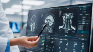
Step Three: Brain Imaging — The Most Important Step
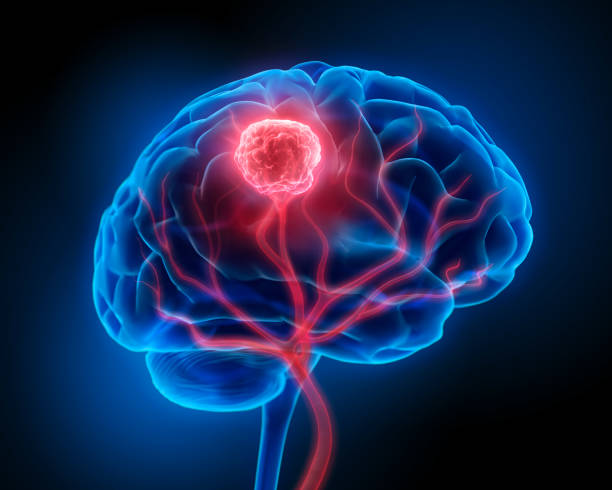
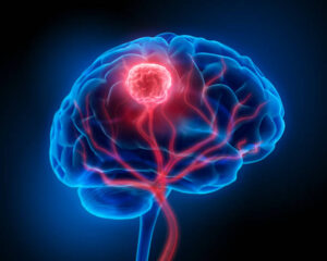
This is where the diagnostic picture really starts to come into focus. Brain imaging is the single most important tool in identifying a brain tumor, and there are two main types used.
MRI — Magnetic Resonance Imaging
MRI is the gold standard for brain tumor diagnosis. It produces extraordinarily detailed images of the brain’s soft tissue, showing the structure in multiple dimensions without using radiation.
Most brain tumor MRIs are done “with contrast,” meaning a substance called gadolinium is injected into the bloodstream through an IV before or during the scan. Gadolinium is a contrast agent that accumulates in areas where the blood-brain barrier has been disrupted — which tumors typically cause. On the scan, these areas appear brighter than surrounding tissue, making the tumor stand out clearly.
The MRI itself involves lying completely still inside a large cylindrical machine while it produces magnetic fields and radio waves to create the images. The machine is noisy — most people describe a combination of loud knocking and buzzing sounds — and the scan can take anywhere from 30 minutes to over an hour depending on what the radiologist is looking for.
The images produced can show the tumor’s location, approximate size, shape, and borders. They can suggest whether it’s pushing into surrounding tissue or remaining well-contained. They can sometimes indicate what type of tumor it might be, based on how it looks on the scan. But imaging alone cannot definitively identify tumor type — that requires tissue analysis.
CT Scan — Computed Tomography
CT scans are faster than MRIs and more widely available, which makes them useful in emergency situations — for example, when someone presents to an emergency room after a seizure and doctors need a rapid look at the brain.
A CT scan uses X-rays taken from multiple angles and processed by a computer to create cross-sectional images of the brain. Like MRI, it can be done with contrast to highlight abnormal areas.
CT scans are good at detecting many brain tumors, bleeding, swelling, and other significant structural changes. However, they provide less detail than MRI for soft tissue evaluation, which is why MRI is preferred when time allows and a brain tumor is specifically suspected.
In many cases, a patient will have a CT scan first — often in an emergency setting — and then follow up with an MRI for more detailed assessment.
Step Four: Referral to a Specialist
Once imaging reveals something suspicious, the next step is an urgent referral to a specialist — typically a neurologist or a neurosurgeon, and often both.
A neurologist specializes in diagnosing and treating conditions of the nervous system. They’ll review the imaging, conduct their own detailed neurological examination, and help coordinate the next stages of diagnosis and care.
A neurosurgeon is a surgeon who specializes in the brain and spinal cord. Even if surgery isn’t ultimately needed, neurosurgeons are often involved early because their assessment of the tumor’s location and characteristics helps determine whether a biopsy is possible and what surgical options might look like.
Some patients will also be referred to a neuro-oncologist — a doctor who specializes specifically in brain tumors and their medical treatment — though this referral often comes slightly later in the process, once a diagnosis has been confirmed.
Step Five: Additional Imaging and Functional Tests
Depending on what the initial MRI showed and where the tumor appears to be located, the medical team may order additional specialized tests before deciding on next steps.
Functional MRI (fMRI)
If a tumor is located near areas of the brain responsible for language, movement, or other critical functions, functional MRI can map exactly which parts of the brain are handling those functions in this specific patient. This information is crucial for surgical planning — it helps surgeons understand what can safely be approached and what must be avoided.
MR Spectroscopy
This specialized form of MRI analysis measures the chemical composition of tissue in and around the suspicious area. Different tumor types have different chemical signatures, which can help narrow down what the tumor might be before a biopsy is performed.
Perfusion MRI and Diffusion Tensor Imaging
These techniques provide additional information about blood flow within the tumor and the pathways of white matter tracts running through and around it — both helpful for planning treatment.
PET Scan — Positron Emission Tomography
PET scans measure metabolic activity in tissue. Cancer cells tend to consume glucose at a much higher rate than normal cells, making them appear as areas of increased activity on a PET scan. While not typically the first imaging tool used, PET can provide useful additional information in certain cases, particularly when trying to distinguish tumor from radiation damage in a patient who has already received treatment.
Step Six: The Biopsy — Confirming the Diagnosis
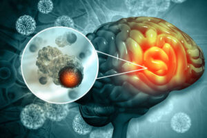
This is the step that confirms everything. Imaging can strongly suggest a brain tumor and give important information about its likely nature, but only tissue analysis under a microscope can definitively identify what type of tumor is present and what grade it carries.
A biopsy involves obtaining a sample of tumor tissue that a pathologist — a doctor who specializes in analyzing tissue — can examine in the laboratory.
Stereotactic Needle Biopsy
This is the most common biopsy method when a tumor cannot be safely removed through open surgery. Using the three-dimensional coordinates mapped by MRI, a neurosurgeon drills a small hole in the skull and inserts a thin needle with precise guidance to collect a sample from the tumor.
The procedure is done under either general anesthesia or local anesthesia with sedation, and most patients are in the hospital for only one to two days afterward. The small hole in the skull heals without the need for plates or major reconstruction.
Surgical Biopsy During Tumor Removal
When imaging suggests that the tumor can and should be surgically removed, the biopsy and the treatment often happen simultaneously. During the surgery, a sample is sent to pathology while the operation continues, sometimes with results coming back within minutes using a process called intraoperative frozen section analysis.
The tissue obtained through either biopsy method then goes through detailed laboratory analysis.
Step Seven: Pathology and Molecular Testing
This is where modern brain tumor diagnosis has become remarkably sophisticated over the last decade — and where the results can make a significant difference to treatment planning.
The pathologist examines the tumor tissue under a microscope, looking at the characteristics of the cells to determine tumor type and grade. But increasingly, this visual analysis is only the beginning.
Molecular and genetic testing of the tumor tissue has become a standard part of brain tumor diagnosis. Specific genetic markers found in the tumor cells can provide critical information about how the tumor is likely to behave and how it might respond to different treatments.
For example, the IDH mutation status of a glioma significantly affects prognosis. Tumors with IDH mutations generally behave less aggressively than those without. The MGMT methylation status affects how well the tumor is likely to respond to chemotherapy with a drug called temozolomide. The presence or absence of a chromosomal deletion called 1p/19q co-deletion helps distinguish between different glioma subtypes with different expected outcomes.
These molecular results can take days to weeks to come back, which is why the full diagnostic picture sometimes takes several weeks to complete even after surgery or biopsy.
Step Eight: The Final Diagnosis and Staging
Once imaging, pathology, and molecular testing results are all available, the medical team brings everything together into a final diagnosis.
This diagnosis includes the specific tumor type — glioblastoma, meningioma, ependymoma, medulloblastoma, and so on. It includes the tumor’s WHO grade from 1 to 4, indicating how aggressive it is expected to be. And it includes the molecular characteristics that shape both prognosis and treatment recommendations.
This complete diagnosis is what guides everything that comes next. Surgery planning, radiation protocols, chemotherapy choices, and monitoring schedules are all built on this foundation.
The Waiting — What Nobody Talks About Enough
My colleague described the diagnostic process not primarily in terms of the tests themselves, but in terms of the waiting.
Waiting for the MRI appointment. Waiting for the MRI results. Waiting for the specialist appointment. Waiting for the biopsy. Waiting for the pathology results. Waiting for the molecular testing. Three weeks of her life lived in the gaps between appointments, with an enormous, unresolved question sitting at the center of everything.
If you’re in that waiting period right now — or someone you love is — I want to acknowledge that it’s genuinely one of the hardest parts of this experience. The uncertainty is often worse than the information, even when the information is difficult.
Two things that can help during the diagnostic process: writing down every question you want answered before each appointment, because it’s easy to forget in the moment, and bringing someone with you to appointments when possible, because a second set of ears catches things that are easy to miss when you’re anxious.
What to Ask Your Doctor During the Diagnostic Process
Having questions ready can help you feel more in control and get more useful information from each appointment.
- What exactly does the imaging show, and what does that mean practically?
- What type of tumor are you suspecting, and why?
- Is a biopsy needed, and what does that procedure involve?
- What molecular testing will be done on the biopsy tissue?
- How long will results take at each stage?
- When will we have enough information to discuss treatment options?
- Who else will be involved in reviewing my case — a tumor board? A neuro-oncologist?
- Should I be getting a second opinion, and would you be comfortable with that?
A good medical team will welcome these questions. If anyone makes you feel that asking questions is a burden, that itself is useful information about whether you’re in the right place.
A Final Word
The diagnostic process for a brain tumor is not a single test and a single answer. It’s a sequence of steps, each one building on the last, that takes days or weeks to complete fully.
Understanding the process before you’re in the middle of it doesn’t make it less frightening. But it does mean you’re not navigating it blind — and that every scan, every specialist appointment, and every waiting period has a place in a process that’s moving you toward answers.
My colleague told me that the hardest thing wasn’t the MRI or the biopsy. It was not understanding what was happening or why. She spent energy she didn’t have on anxiety that came from confusion, not from the information itself.
You deserve to understand the process you’re going through. That understanding won’t change what the results say — but it can change how you carry the wait.
Disclaimer: This article is written for educational and informational purposes only and does not constitute medical advice. Diagnostic processes vary between individuals and medical institutions. Please consult a qualified neurologist or neurosurgeon for guidance specific to any individual situation.